 |
| HZO |
What
is herpes zoster ophthalmicus (HZO)?
It is a viral disease
that involves skin of head and eyes and involves the ophthalmic branch of
trigeminal nerve (CN-5).
What
causes herpes zoster ophthalmicus?
It is caused by
varicella-zoster virus, which is the same virus that causes chicken-pox. The
infection occurs when the latent herpes zoster virus in the neurosensory
ganglions is reactivated. Old age, weakened immune system (HIV), emotional or
physical stress and fatigue precipitate the reactivation of varicella-zoster
virus.
What
are sign and symptoms of herpes zoster ophthalmicus?
Caused by a virus herpes
zoster, HZO presents with flu-like illness, low grade fever, headache, malaise,
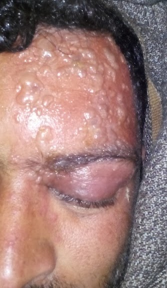
maculopapular rash over the head and blepharoconjunctivitis. However, vesicular
rash over the dermatome of first division of cranial nerve 5 (CN5-V1) is the
hallmark of HZO. Inflammation, edema and ptosis of upper eye-lid are commonly
seen.
What
tests should be carried out for herpes zoster ophthalmicus?
Most of the time, no tests are needed as clinical findings are diagnostic. However, some tests can be carried out to confirm or exclude the diagnosis:
- Antigen detection by immunofluorescence
- Viral swabs of vesicle for culture
- VZV DVA PCR
What is the treatment of herpes
zoster ophthalmicus?
Antiviral therapy is
the main stay of management of HZO.
For
systemic illness:
Tablet Acylex
(Acyclovir) 800 mg – Take one tablet five times a day for seven days.
[Acyclovir should be administered within 3 days (72 hours) of the presentation of ophthalmic features. After 72 hours, the effect of acyclovir is diminished.]
For
topical use:
Acylex Ointment (Topical
antiviral ointment) – Apply over the affected areas for two times a day.
Fusiderm ointment (Antibiotic/antibacterial
ointment) – Apply over the affected areas three times a day.
For
eye care:
Lequix eye drops
(contains levofloxacin) - One drop four times a day for 20-30 days
Sanitovir (antiviral
eye ointment) - Apply four times a day for 20-30 days.
Cold compresses –
relieves pain
Lidocain cream 5% -
topically used to relieve pain
NSAIDS (conventional pain-killers)
– Intramuscular or oral use
What
are the complications of herpes zoster ophthalmicus?
If timely management is
not offered, HZO can lead to serious complications like:
- Epithelial, stromal,
and disciform keratitis (persistent vasculitis may lead to eurotrophic
keratitis, mucus plaque keratitis and lipid degeneration of corneal scars)
- Dry eye
- Anterior uveitis
- Necrotizing retinitis
- Cranial nerve palsies
(usually facial nerve; however, oculomotor (CN-3), trochlear (CN-4), and
abducens (CN-6) nerve palsies may also occur
- Lagophthalmos
- Postherpetic neuralgia
(PHN)
- Raised intraocular
pressure
- Orbital apex syndrome
- Vision loss
- Optic neuritis [especially retrobulbar optic neuritis- if optic neuritis occurs, methylprednisolone (Solu Medrol) 1000mg intravenous or intramuscular for 3 to 5 days plus prednisolone (Deltacortil) 60mg per day for 10 days are advised along with antiviral therapy and other measures].
How
to prevent herpes zoster infection?
The patient with active
varicella-zoster infection should avoid contact with susceptible persons such
as premature infants and immunocompromised persons, until the lesions/vesicles
are crusted.
Varicella-zoster
vaccine (attenuated VZV) is safe, effective and well-tolerable to reduce the
subsequent attacks and decrease the morbidity and mortality of zoster virus. It
is a lyophilized preparation of attenuated VZV. A single dose of 0.65 ml is
injected subcutaneously in the region of deltoid muscle. Each 0.65 ml of zoster
vaccine contains 19400 PFU (plaque-forming-unit) of Oka stain of VZV. Do not inject intravenously or
intramuscularly. Booster dose of zoster vaccine is not recommended.
Reference:
-
Sanjay S, Huang P, Lavanya R. Herpes zoster
ophthalmicus. Curr Treat Options Neurol 2011;13(1):79-91.
-
Shaikh
S, Cristopher N. Evaluation and management of herpes zoster
ophthalmicus. Am Fam Physician 2002;66(9):1723-30.
- Gelb LD. Preventing
herpes zoster through vaccination. Ophthalmology 2008;115(2 Suppl):S35-8.
A Case Summary of the patient with the same condition
Name:(Patient)
Date of birth: 15/03/1977 (Age: 37 years)
Gender: Male
Residence: Punjab,
Pakistan
Medical Summary:
No improvement was observed. The
patient was advised to have CT scan brain and right eye visual field. Both CT
scan brain and right eye visual field were normal.
Final Diagnosis: Post
Herpes Zoster Ophthalmicus (HZO) Retrobulbar Optic Neuritis of left side,
leading to complete left loss of vision
Clinical and Treatment Summary:
Mr. Muhammad Saeed experienced severe pain behind the left eye and small sized
vesicular rashes over the left side of forehead on February 17, 2014. The local
doctor took it for some allergy and gave some anti-allergic drugs. The rashes
increased in size spreading around the left eye. Left eye was swollen and
closed. A skin specialist and an ophthalmologist were consulted on 19/02/2014. Diagnosis
of Herpes Zoster Ophthalmicus (HZO) was made. The skin specialist put
the patient on the following drugs:
Acylex 800 mg
x P/O x 5 times a day (for 10 days)
Acylex Ointment
x BD
Fusiderm ointment
x TDS
The
eye specialist advised no drug as eye examination was normal.
On
24/02/2014, rashes subsided but the patient experienced vision loss and
immediately was examined by the eye specialist and the diagnosis of HZO Retrobulbar Optic Neuritis of left
side, leading to complete left loss of vision was confirmed. The
patient was put on the following drugs in addition to the above mentioned
medication:
Lequix eye drops
x 4 times a day (for 30 days)
Sanitovir x
4 times a day (for 30 days)
Deltacortil 5mg
x 5 x TDS (continued for 3 days)
The unsatisfied patient attended
another eye specialist on 27/02/2014. And, he got the following treatment:
Solu Medrol
1000mg
x IV (in 1000ml N/S) x (for 3 days)
Deltacortil
60mg
x OD (for 10 days)
Keywords: Herpes zoster ophthalmicus, HZO, Trigeminal nerve, Varicella-zoster virus, Keratitis, Blepharoconjunctivitis, Vision loss, Antiviral therapy, Acyclovir. Neurosensory ganglion